
Released 8/5/26
You can use artificial intelligence to detect possible caries, calculus, periapical radiolucency (PRL), and notable margins on intraoral X-ray images in Dentrix Ascend Imaging. With an Accelerate subscription, Pearl AI is available at no additional cost. With an Essentials or Pro subscription, Pearl AI is available as an add-on that must be purchased separately.
Automatic Submission for Analysis
After you acquire a single-image exam (such as one bitewing) or the last image in a multi-image exam (such as a full-mouth series), when the image for the exam or the first image of the exam is loaded in the viewing area, the image is submitted automatically for analysis.
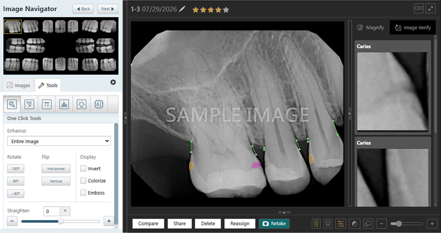
When you view any image in an exam, the image is submitted automatically for analysis.
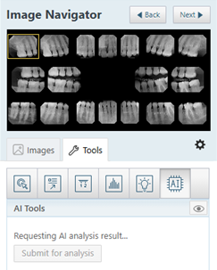
Showing and Hiding Findings
1. While you are viewing an image, on the Tools tab, click the AI tool.
The findings appear.
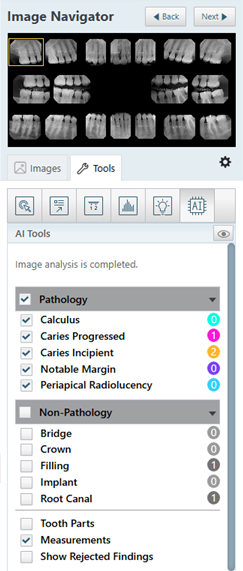
Note: For a given finding, the number of instances found in the image being viewed appears in the circle to the right of the finding name.
2. Select the checkbox of each finding that you want to show. Clear the checkbox of each finding that you want to hide.
The following options are available:
Pathology:
Calculus – A shaded teal box appears.

Caries Progressed – A shaded pink area appears.

Caries Incipient – A shaded yellow area appears.

Notable Margin – A shaded purple box appears.

Periapical Radiolucency – A shaded blue area appears.

Non-Pathology:
Bridge, Crown, Filling, Implant, or Root Canal – A shaded gray box appears.

Other:
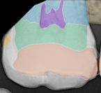
Tooth Parts – Multiple shaded colored areas appear.
Measurements – Colored lines appear.

Rejected Findings – Select this checkbox if you want to view findings that were rejected.